
X-Ray
How does X-Ray work?
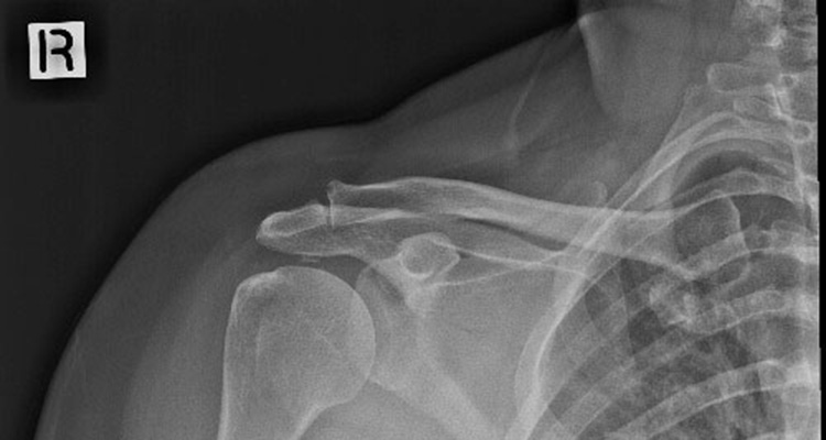
A radiograph is an X-ray image obtained by placing a part of the patient in front of an X-ray detector and then illuminating it with a short X-ray pulse. Bones contain much calcium, which due to its relatively high atomic number absorbes X-rays efficiently. This reduces the amount of X-rays reaching the detector in the shadow of the bones, making them clearly visible on the radiograph. The lungs and trapped gas also show up clearly because of lower absorption compared to tissue, while differences between tissue types are harder to see.
Radiographs are useful in the detection of pathology of the skeletal system as well as for detecting some disease processes in soft tissue. Some notable examples are the very common chest X-ray, which can be used to identify lung diseases such as pneumonia, lung cancer or pulmonary edema, and the abdominal X-ray, which can detect bowel (or intestinal) obstruction, free air (from visceral perforations) and free fluid (in ascites). X-rays may also be used to detect pathology such as gallstones (which are rarely radiopaque) or kidney stones which are often (but not always) visible.
“The staff were really pleasant and I was surprised how fast and easy the process was.”
Preparation for X-Ray
In most cases, no specific preparation is required. Your referring physician or specialist will confirm this with you. Depending on the location of the X-ray, we may ask you to change into one of our gowns and/or remove items that could interfere with the image. Our Radiographer will show you where to change and store your belongings. You may bring a support person if you need assistance with mobility or moral support. Parents may sit with their children. Important – You must inform us if there is a chance you may be pregnant. Many exams will not be performed due to radiation risk to unborn foetuses. If your X-ray is necessary then precautions to reduce risk can be taken.
During
Please allow up to 20 minutes for your X-ray. For a clear image you must remain still as directed. Be assured that the exam is carried out by an experienced and caring technician who will talk you through the process, position you carefully (either sitting, standing or lying down) and make sure you are comfortable.
After
Images will be reported directly to your physician within hours (sometimes minutes) of being taken. If the cost of your X-ray is to be covered by ACC, there will be a surcharge which is payable on the day of your exam.